Optimising the delivery of stroke care
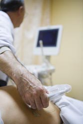
In patients with acute cerebral ischemia, brain perfusion can be analysed by means of different diagnostic techniques, including computed and magnetic resonance tomography among others. However, it is the continuous development of non-invasive ultrasound techniques that has provided invaluable clinical applications for the assessment of intracranial arterial diseases. Ultrasound can be utilised at the bedside for critically ill patients and provide timely information on arterial dissection and occlusion of brain arteries, as well as embolic occlusion. The concept of ultrasound perfusion analysis was not new; but the ability to estimate perfusion parameters with accuracy has only been made possible with recent developments in ultrasonographic imaging techniqes. Localisation of specific biochemical epitopes with targeted contrast agents afforded the opportunity for imaging thrombus material in acute vessel occlusions. It also provides an effective means for the detection of micro-embolic signals. Moreover, software development at the INSERM laboratories in France has allowed rapid electronic transfer of data from the detector arrays and rapid image reconstruction for subsequent perfusion analysis. Studies were undertaken to ascertain the optimal delivery mode of microbubbles with antibodies to activate platelets for targeted imaging, and the results were integrated into the image analysis software. The new software supports the automatic factor analysis of medical image sequences (FAMIS), along with the conventional parametric analysis of temporally arranged sequences. To minimise respiratory motion artefacts in ultrasound contrast imaging sequences, and furthermore to improve parametric and FAMIS imaging from time sequences, dedicated software developed in MATLAB was added. The software package, readily available on a CD-ROM, can be implemented either step-by-step under the permanent supervision of the user or automatically both for clinical and pharmaceutical research applications.


