Realistic models give a clearer view of breast cancer
It is estimated that 1 in 11 women in the EU will develop breast cancer before the age of 74. Although breast cancer mortality rates have been declining in most EU countries, it still has the distinction of being the deadliest form of cancer for women – meaning there’s room for improvement. “One of the best ways to reduce cancer-related deaths is early detection, and that starts with breast imaging,” says Kristina Bliznakova, a biomedical engineer at the Medical University of Varna in Bulgaria. But before a new breast imaging modality can get to work detecting cancer, it first needs to undergo extensive testing to prove its effectiveness and safety. However, due to ethical considerations, regulations and costs, such experiments cannot be performed on human patients. Instead, researchers depend on physical and virtual models of breast tissues. “Having access to realistic and accurate physical and computational anthropomorphic breast models is an ultimate requirement for the successful development of advanced imaging techniques,” adds Bliznakova. Delivering those models is the EU-funded PHENOMENO project.
Realistic breast models
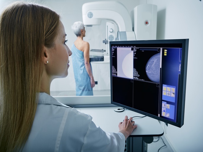
Bringing together industry partners and universities, the PHENOMENO project used actual MRI breast images to deliver innovative computational and physical breast models. “Developing a realistic-as-possible model required our team to conduct extensive research on breast tissue characterisation, breast compression techniques, and the use of breast imaging simulations,” explains Bliznakova. The project, which received support from the Marie Skłodowska-Curie Actions programme, also developed novel printing materials and 3D printing techniques for producing realistic breast models. According to Bliznakova, the resulting virtual and physical models are now ready to be used in the testing of new X-ray-based breast imaging techniques. “We produced an anthropomorphic breast model that’s being used in the testing of a very promising, contrast-enhanced mammography procedure,” she says.
Photon-counting and artificial intelligence
By enabling the effective testing of advanced imaging modalities, the PHENOMENO models represent an important step in reducing the incidence and impact of breast cancer. Yet, as Bliznakova points out, there’s still plenty more work to do. “Because of the strong collaboration established between the project’s partners, we are able to continue our work and further develop our breast models,” she notes. For example, researchers are currently looking to manufacture breast models that feature lesions. The models will then be used to test a new micro-CT scanner being developed by the Medical University of Varna. Furthermore, several of the project’s partners are working on an advanced photon-counting detector, while another is leveraging its expertise in artificial intelligence to help radiologists better interpret breast images. “Not only is this work contributing to the development of advanced techniques and tools, they are also building a community of high-quality researchers equipped with the skills needed to further improve breast cancer diagnostics,” concludes Bliznakova.
Keywords
PHENOMENO, cancer, breast cancer, breast imaging, breast models, MRI, 3D printing, X-ray, mammography, artificial intelligence, radiologists