

Nanoprobes assist prompt detection of gastrointestinal cancer
White light endoscopic surveillance is the method most often used for detecting and removing dysplastic lesions in the gastrointestinal tract. However, the most clinically relevant lesions have a subtle appearance, and there is a high risk of missing them. Therefore, there is an unmet need for novel approaches that can detect premalignant gastrointestinal tract lesions with high sensitivity and help prompt an accurate diagnosis of malignant progression.
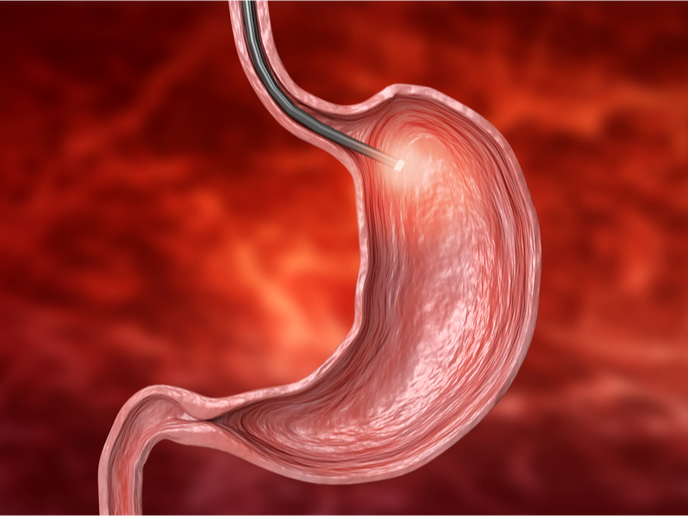
Nanoparticle-based dyes for high-precision coloured endoscopy
The THERAPROBES project has developed a novel chromoendoscopy approach for the improved detection of lesions. “Our goal was to develop dyes that enhance macroscopic structural features and provide contrast of the healthy tissues surrounding the lesions,” explains project coordinator Alan Chan. The research was undertaken with the support of the Marie Skłodowska-Curie Actions programme and involved the development of biodegradable fluorescent silica nanoparticles, also known as Theraprobes. These probes incorporate near-infrared fluorescent dye into a silica-based nanoparticle matrix of 100 nm in size. Theraprobes enabled the endoscopic detection of dysplastic lesions in a mouse model that closely recapitulates oesophageal carcinogenesis. Moreover, following systemic administration in a mouse model of colorectal cancer, Theraprobes facilitated the imaging of the intestinal burden of polyps and adenocarcinomas with high sensitivity, specificity, and precision. Adenomas as small as 0.5 mm2 could be detected throughout the intestinal tract of animals.
Advancement in the clinical implementation of chromoendoscopy
“The most significant achievement of the project was the development of a preclinically viable strategy for improving the diagnostic accuracy of endoscopy and facilitating the early detection of premalignant lesions of the gastrointestinal tract,” emphasises Chan. Because of their size, these fully biodegradable fluorescent silica probes have the potential to extravasate or escape from the blood vessel and accumulate in the tumour without requiring active targeting. As a result, they can serve as ubiquitously applicable imaging agents, while the proposed approach is fully compatible with existing clinical practices and instrumentation. Additional advantages of Theraprobes include the low cost as well as the ease and time of administration. Conventional dyes require a laborious procedure and prolonged examination, which prohibit the widespread use of chromoendoscopy. Theraprobes mitigate the high miss-rate and low diagnostic accuracy of conventional white light endoscopy and negate the perceived drawbacks of chromoendoscopy.
THERAPROBES advantages and prospects
Implementation of Theraprobes will undoubtedly improve the detection rates of premalignant lesions with important implications for the surveillance, management and prevention of colorectal cancers. It will assist the precise resection of stained lesions, avoiding random biopsies, which is the standard practice. Moreover, the prompt endoscopic detection of premalignant lesions will help avoid aggressive interventions, such as colectomy for managing dysplasia in high-risk patients. Significantly, augmented endoscopy using Theraprobes will help direct treatment to early lesions that are amenable to therapeutic interventions. Chan is confident that additional funding is necessary for clinical validation of these nanoprobes and will help the further functionalisation to enable image-guided photodynamic therapy as a first-line therapy or after tumour resection to reduce recurrence risk.
Keywords
THERAPROBES, endoscopy, gastrointestinal tract, chromoendoscopy, nanoprobes, fluorescent silica nanoparticles, colorectal adenomas