

Microwave for breast cancer detection
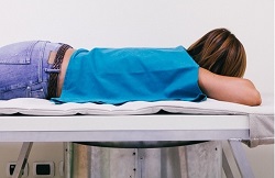
Mammography, the gold standard for breast screening and breast cancer diagnosis, utilises X-ray ionising radiation to generate images of breast tissue. However, it is not recommended for women under 50 years old or during pregnancy due to safety concerns related to ionising radiation exposure. This means that nearly 40 % of women in Europe between the ages of 25-49 cannot use the most conventional breast cancer screening modality. The EU-funded MammoWave project developed a novel procedure that uses microwave frequencies to examine breast tissue. The idea was to perform the first screening using the MammoWave device and then refer positive cases to traditional mammography, thereby limiting the use of harmful radiation. Microwaves in breast screening “Microwave imaging has received increasing attention in the last decades for breast cancer detection due to the considerable difference in the dielectric properties of malignant and normal tissues at microwave frequencies,″ explains project coordinator Dr Gianluigi Tiberi. The MammoWave microwave device consists of a cup that holds the breast when the patient lies on the examination table. There are two antennas, which move around the cup, for irradiating and capturing the microwaves scattered by the breast tissue. The signals measured by the receiving antenna are then processed by specialised software to produce images that represent intensity maps of the breast tissue dielectric properties. A specific algorithm identifies microwave images of non-healthy breasts, which exhibit a greater ratio of maximum to average intensity than normal tissue. “MammoWave is based on an innovative image reconstruction algorithm that processes the microwave signals, thereby detecting the presence of any suspected tumour in the breast tissue,″ continues Dr Tiberi. However, as the breast comprises non-homogeneous regions, a certain level of mismatch can also be seen in the healthy breasts and should be related to tissue anatomy on an individual basis. Clinical application of the MammoWave apparatus Preliminary data on patients and healthy volunteers have resulted in a 91 % sensitivity of the MammoWave apparatus. The accuracy of the imaging device for breast cancer detection is currently being tested in a feasibility clinical study at Perugia and Foligno Hospitals in Italy. MammoWave researchers are confident that their technology is effective and safe for a widespread breast screening approach. As there is no ionising radiation, it is safe to use at any age, in any condition with no limit on frequency of breast screenings. What’s more, in contrast to traditional mammography units that compress the breast, the MammoWave device is more comfortable for the patient. The apparatus can be used in hospitals, clinics and private practices, offering an enhancement in breast screening and breast cancer detection. In conjunction with traditional mammography, it can help decrease false-positive and false-negative results, minimising distress for women. As Dr Tiberi states, “the next step is to start the commercialisation of the medical device, first in Europe and then worldwide.″ He is confident that implementation of MammoWave microwave screening will not only improve healthcare quality but also help cut down healthcare costs.
Keywords
MammoWave, microwave, breast cancer, mammography, ionising radiation