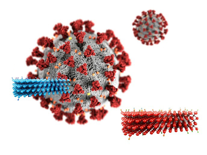
The dynamic potential of immune-safe 2D materials in biomedical applications
Two-dimensional (2D) materials have emerged as powerful tools in biomedical research due to their unique structural and functional properties. They are usually structured as thin layers, exhibiting exceptional surface area, high electrical conductivity, mechanical strength and tuneable chemical characteristics. Moreover, they are biocompatible and capable of interacting with biological molecules and cells. Collectively, these properties make 2D materials highly versatile and adaptable for a range of health-related applications, from targeted drug delivery and biosensing to tissue engineering and regenerative medicine.
MXene immune system interaction
Undertaken with the support of the Marie Skłodowska-Curie Actions programme, the SEE project focused on MXenes, which are 2D structures consisting of carbon and transition metals with remarkable biomedical potential. MXenes can modulate and interact with the immune system depending on the application. This means that they could be used to promote beneficial immune activation, for example, in wound healing where immune cells play a vital role in tissue repair. Conversely, for conditions requiring immune suppression, MXenes might help alleviate inflammation. These dual capabilities of MXenes, however, raise concerns about harmful inflammatory responses or other adverse immune effects. “Our goal was to investigate how MXenes interact with immune and skin cells, so as to design safe materials for clinical use,” explains project supervisor Lucia Gemma Delogu.
Advanced techniques for MXene safety evaluation
The SEE project utilised a range of sophisticated techniques to assess MXene interactions. Ex vivo and in vivo assays with primary immune cells and human skin models provided essential data on cytotoxicity, immune cell activation and cytokine release. To achieve precise, high-resolution results, the project employed single-cell mass cytometry by time of flight CyTOF, which leverages metal-labelled antibodies for analysing multiple cellular markers and performing immune cell profiling. Researchers combined CyTOF with ion-beam imaging by time of flight (MIBI-TOF) to visualise MXenes within tissues and determine their distribution and cellular interactions. By pioneering the use of CyTOF and MIBI-TOF in a label-free strategy named LINKED, the SEE project has set a new standard for high-throughput single-cell analysis and for evaluating biocompatibility of 2D materials. This approach allowed researchers to examine MXene interactions across a spectrum of cell types and tissues, offering a unique perspective on the potential of MXenes in a clinical setting. MXenes exhibited biocompatibility in skin applications and promoted wound healing, without compromising the viability of human epidermal keratinocytes. This lack of cytotoxicity is central for preserving skin health in cosmetic and biomedical applications.
Strategy importance and future directions
“The insights generated by LINKED not only enhance our understanding of MXenes in biomedicine but also contribute to designing 2D materials with tuneable immune properties,” highlights Delogu. The project has garnered interest from industry partners, and preliminary findings on MXene safety have led to a provisional patent application, marking a critical step toward commercialisation. Importantly, the multiplexed LINKED technique can be used for the detection of other 2D materials in biomedical research as well as in applications beyond healthcare in fields like catalysis, energy storage and artificial intelligence.
Keywords
SEE, MXene, 2D materials, CyTOF, immune cells, MIBI-TOF, skin cells


