A new era in chromosome research
Traditionally, chromosome investigation involves the isolation and preparation of metaphase chromosomes from mitotic cells. However, this process is unsuitable for in vitro experiments as the chromosomes are fixed, prohibiting the study of their dynamics.
A microfluidics device for isolating and studying chromosomes
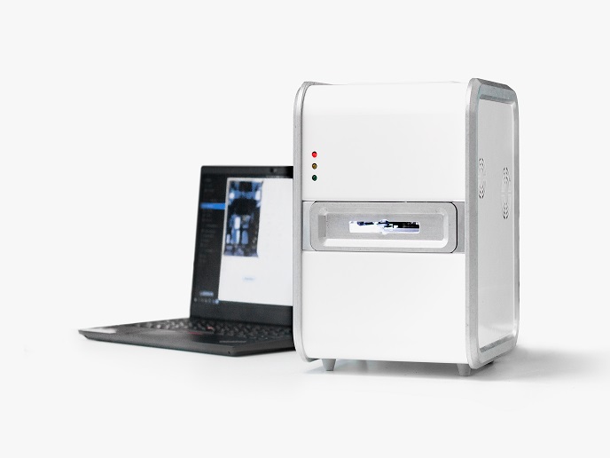
To address this issue, the EU-funded CHROMAVISION project developed a pioneering chromosome imaging and manipulation platform that allows the investigation of individual chromosomes from cell samples using a super-resolution microscope. “Our instrument allows for the first time the detailed study in real time of metaphase chromosome structure and their associated proteins,″ explains project coordinator Prof. Gijs Wuite. The CHROMAVISION instrument combines lab-on-a-chip microfluidics, multiple-trap optical tweezers and super-resolution fluorescence microscopy. Single chromosomes can be captured by attaching small micron-sized beads to the ends of the metaphase chromosome and holding these beads in two optical tweezers. These beads serve to hold the chromosome at a fixed position in the microfluidic device chamber and can even stretch the chromosomes by moving the beads apart. Additionally, the instrument provides the capacity to use laser beams to scan chromosomes, thereby providing information on the position of associated proteins that have been labelled with fluorophores. By combining images of the positions of several key proteins that build the scaffold of a metaphase chromosome, scientists can obtain a 3D map of how the chromosome is constructed. Project partners have generated a production-ready prototype, which they shipped to various labs for testing purposes. The prototype allows super-resolution molecular imaging of the DNA in the chromosome with the capacity to test the elasticity of a metaphase chromosome as well as to visualise specific proteins within the chromosome.
CHROMAVISION advantages and future prospects
According to Prof. Wuite, the biggest achievement of the project was “the development of a single instrument capable of isolating, holding and imaging metaphase chromosomes in cells with super-resolution.″ He emphasises that this wouldn’t have been possible without the hard work and successful collaboration of the five different research teams from three countries. The biggest advantage of the CHROMAVISION instrument compared to existing solutions is its capacity to utilise non-fixed metaphase chromosomes for imaging and manipulation studies. This allows researchers to look at the dynamic aspects of chromosomes such as protein motion and/or stretching of the chromosomes. It also facilitates the imaging of DNA protein interactions at the molecular level in real-time under near-physiological conditions. Prof. Wuite is confident that the CHROMAVISION platform will push the boundaries of what is currently possible in microfluidics and super-resolution microscopy and fuel the next decades of chromosome studies. The instrument will help address key challenges in clinical and fundamental chromosome research, potentially resulting in breakthrough discoveries. Better imaging and understanding of the chromosomal mechanisms will contribute to our knowledge of the aetiology of human diseases and aid drug discovery. From a clinical perspective, the CHROMAVISION instrument is expected to facilitate the diagnosis and monitoring of cancer heterogeneity. Partners have secured future funding to improve chromosome analysis output that is suitable for patient sample analyses.
Keywords
CHROMAVISION, metaphase chromosome, microfluidic, beads, super-resolution, microscopy, cancer, dynamic, optical tweezers, prototype, fluorophore


