Breast cancer detection boosted in Bulgaria via models
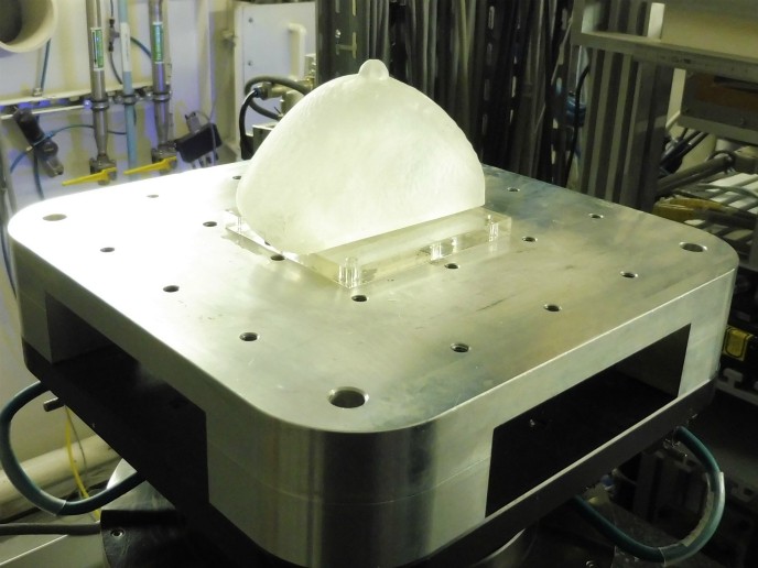
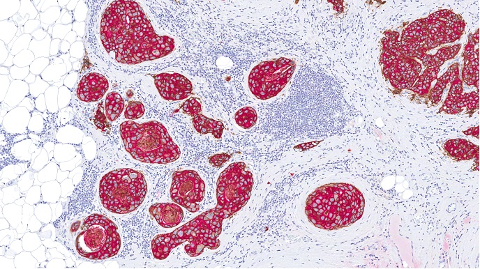
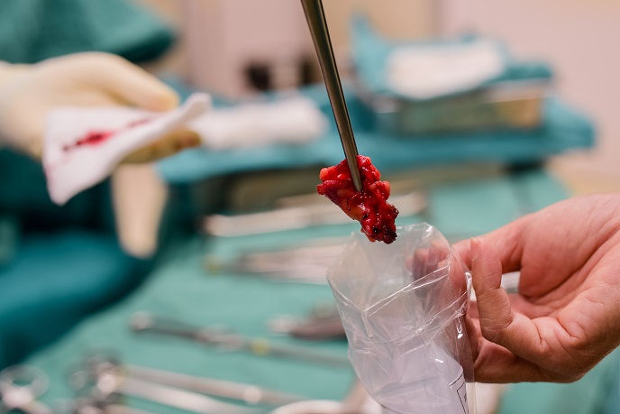
Breast cancer is the scourge of our time. It affects one in eight women in Europe at some point in their lives. Approximately 20 % of breast cancers occur in women under the age of 50. Early diagnosis can greatly improve recovery and survival. The EU-funded project MaXIMA focused on creating 3D computational models of malignant breast tumours. These can be used to develop new techniques for detecting more complex forms of breast cancer that are difficult to diagnose with standard examination methods. Formulated in partnership with the Catholic University of Leuven in Belgium and the University of Naples Federico II in Italy, MaXIMA’s innovative research has the potential to save many lives. Unmasking breast cancer in dense breasts The research conducted by MaXIMA focused on enhancing techniques to detect small and irregular-shaped tumours in dense breast tissues. “Despite recent technological advances such as digital mammography, detecting cancers hidden in dense breast tissues is challenging,” says project coordinator Kristina Bliznakova. “Unfortunately, contrary to expectations, breast cancer mortality continues to rise in many EU countries, and one of the most important reasons could be related to the limitations of current technology to screen dense breasts.” Breast tomosynthesis is a newer advanced technology that has been designed to overcome the limitations associated with conventional 2D mammography. The examination uses low-dose X-rays, and can be carried out at the same time as the 2D mammogram. Images are taken at varying angles and reconstructed using a computer into thin slices of 3D volume. These thin slices enable radiologists to detect small breast tumours that are masked by the overlying gland tissue. Another X-ray imaging technique that can add more information on tissue structure is phase-contrast X-ray tomography. Traditional mammography relies on the decrease of the X-ray beam's intensity when traversing the body tissues. However, phase-contrast X-ray imaging measures the difference in the way an X-ray beam oscillates through normal tissue compared with denser tumour tissues. The technique provides a sharper picture of subtle changes in tissue density as it increases the visibility of thin edges and border details in many specimens. 3D models to better outline the tumour Despite their uptick in popularity, advanced X-ray breast imaging techniques need to be complemented by computer models and simulations to be entirely effective in practice. “In most cases, tumours form inhomogeneous masses with no distinct boundaries. This prevents us from clearly distinguishing the contours of the cancer formations,” explains Bliznakova. “Three-dimensional computational and physical models are powerful tools in the hands of engineers, physicians and physicists. Advanced models help scientists accurately define breast shapes, glandular tissue distribution and cancer mass shape and type,” adds Bliznakova. In light of this, project researchers developed novel computational models of hard-to-diagnose breast tumours such as those surrounded by dense parenchyma. Furthermore, they created physical anthropomorphic models, known as phantoms, of breasts and tumours for testing and validating X-ray imaging techniques including breast tomosynthesis and phase-contrast imaging. Suitable 3D printing techniques and materials were investigated for producing physical breast phantoms. Close cooperation between the partnering institutions helped to significantly increase the scientific and technological capacity of the Technical University of Varna in the X-ray breast imaging field. Innovative projects like MaXIMA should help establish Bulgaria as a hub for research and development in Europe.
Keywords
MaXIMA, breast cancer, X-ray imaging, mammography, Bulgaria, Technical University of Varna, dense breast tissue, 3D model


