New insight into the development of new blood vessels could lead to improved cancer therapies
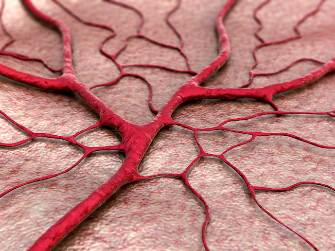
Angiogenesis is the process of formation, growth and stabilisation of new vessels, originating from the pre-existing vascular network. It is involved in vascular network formation during development but also in homeostasis, the ability to maintain internal stability in an organism to compensate for environmental changes. Sprouting angiogenesis is one way in which an endothelial cell, (i.e. the cells that line and form all blood vessels), adapts its shape to become invasive. It does so by extending protrusions in the direction of the future sprout and starts to migrate in this direction. This cell will lead the way for the following cells that will make the building blocks of the forming vessel. The MTUB-ANGIO project’s goal was to investigate the role of the microtubule cytoskeleton, a network of scaffolding tube-like fibres in blood vessel formation. As principle investigator, supported by the Marie Curie programme, Dr Maud Martin explains, “In a cell, microtubules can be organised through anchoring at the centrosome, the main place where cell microtubules may be organised. If they aren’t organised there then a protein named CAMSAP2 binds to one end of the microtubules and stabilises them.” The centrosome was generally believed to play an important role in cell migration by acting like a compass, pointing in the direction that the cell will move. To challenge this view, the project used in vitro culture of endothelial cells, isolated from the vessels of human umbilical cords. These they cultured either as 2D sheets, to monitor cell migration in wound-healing assays, or as spheres embedded into 3D collagen gels and forming sprouts. The work is set out in a recently published paper. “We used a drug to target the centrosome to show that the microtubules anchored at the centrosome are not required for endothelial cell migration and sprouting. In contrast, removing the non-centrosomal microtubules by silencing CAMSAP2 prevents the cell from migrating in a directional way. It also causes the formation of sprouts in 3D matrix to be destabilised.” This in-depth understanding of the mechanisms behind endothelial cell migration and sprouting is significant. As angiogenesis is implicated in several disorders, fundamental understanding of its molecular and cellular mechanisms is crucial for developing innovative therapeutic treatment in a long-term perspective. “Angiogenesis is intimately linked to cancer development as tumours need a blood supply to grow and disseminate. Microtubules are already a target for cancer therapy, so the knowledge coming from this project is a good opportunity to optimise the use of existing drugs in the context of angiogenesis-based therapies,” says Dr Martin. She believes the project was a success because she was able to combine the knowledge and cutting-edge imaging methods she successfully learnt in the laboratory under her supervisor Dr Anna Akhmanova, with her former experience and expertise. “By being able to look at detailed cellular mechanisms during 3D physiological processes, including animal models, I have played a part in bridging the gap between information coming from in vitro biology and its translation in vivo.”
Keywords
MTUB-ANGIO, angiogenesis, cellular mechanisms, microtubules, microtubule cytoskeleton, cell migration, vascular network, endothelial cell


