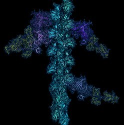
Shedding new light on the workings of motor proteins
Molecular motors are remarkable biological molecular machines that are the essential agents of movement in living organisms. These tiny machines harness the chemical-free energy released by the hydrolysis of ‘adenosine triphosphate’ (ATP) to perform mechanical work such as muscle contractions, cell motility and cell division. Scientists within the RMPSHSSI (Revealing myosin’s power stroke with high-speed scattering interferometry) project recorded the motion of myosin 5. These proteins function just like nanoscale lorries might, travelling remarkably long distances while carrying a cargo. They also look like two-legged creatures that take very small steps. The team used a new optical microscopy technique, called interferometric scattering microscopy, that can see tiny steps of tens of nanometres captured at up to 1 000 frames per second. This powerful imaging technique overcomes challenges associated with the limited resolution of most optical microscopes as well as the quick movement of molecules. Using this technique, the team obtained timing and spatial information about the movement of myosin 5 along a fibrous track. These molecular motors produced motion in a mechanical step known as a power stroke. Project findings shed further insight into how cells function, while also helping step up efforts aimed at building efficient nanomachines. Use of this new optical tool will facilitate a better understanding of cell transport as well as cell division, replication and communication.
Keywords
Motor proteins, stiff-legged, actin filament, interferometric scattering microscopy, cell transport


