'Looking into' cell differentiation
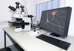
Patients have a host of previously unavailable possibilities thanks to in vitro generation of hepatocytes to treat liver failure or cardiomyocytes to address acute coronary disease. The 'Differentiation dynamics in size-controlled embryoid bodies' (DIFFEBIMG) project focused on embryoid bodies (EBs), 3D aggregates of differentiating embryonic stem cells. Scientists developed two types of growth assays for EBs, one using microfluidic devices and another in microwell arrays. Microfluidic devices allowed for monitoring interactions between the isolated pairs of cells. Formation of uniform-size EBs containing patches of cardiomyocytes took place within the microwell arrays. Growth and differentiation were observed for over two weeks. A two-photon laser scanning microscopy system helped to unravel the relation between the signalling events and differentiation decisions using fluorescent embryonic stem cell markers. Scientists conducted several live imaging experiments to monitor the effect of focal signalling on early mesodermal differentiation. The developed system for wild type and interventional monitoring and analysis of spatiotemporal developmental changes in 3D tissue is a unique tool. Study of differentiation and signalling events will help to elucidate connections between signal and cell fate.
Keywords
Cell differentiation, in vitro differentiation, embryonic stem cells, cardiomyocytes, embryoid bodies


