Improved image analysis for MRI
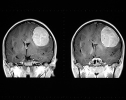
Medical imaging is a crucial tool in clinical environments, particularly in developing image-based quantitative tools as part of cancer therapy and tumour response measurement after therapy. The 'Medical image analysis for cancer treatment monitoring and tumor atlas formation' (MICAT) project worked on the development of new medical computation methods and a medical image analysis framework for monitoring during cancer treatment. In particular, MICAT focused on tumour response assessment and formation of a novel probabilistic brain tumour atlas. Tumour segmentation from imaging data is challenging due to the high diversity in appearance of tumour tissue between patients and, in some cases, with normal tissue. The MICAT team developed a new segmentation method based on the cellular automata (CA) algorithm. Validation studies showed that the final algorithm outperformed graph cut and grow cut algorithms with a lower sensitivity to initialisation and tumour type. Image registration applications include combining images of the same subject from different modalities and image guidance during interventions. MICAT researchers developed a new rigid registration method based on anatomical brain landmarks to compare baseline and follow-up image volumes from a brain tumour patient. The new technique involves serial contrast-enhanced T1 weighted magnetic resonance (MR) images. T1-weighted scans depict the differences in the spin lattice relaxation time of the tissue. The new protocols mean that tumour volume and diameter change measurements can be carried out with increased accuracy and reliability. Furthermore, MICAT are looking into new local criteria using the Lagrange deformation tensor. To date, seven papers have been published detailing the accomplishments of the MICAT project. As the software tools will be made available in clinical practice, there will be increased efficiency in tumour radiosurgery and analysis of brain tumour response on follow up after treatment.


