Shedding more light on the human brain
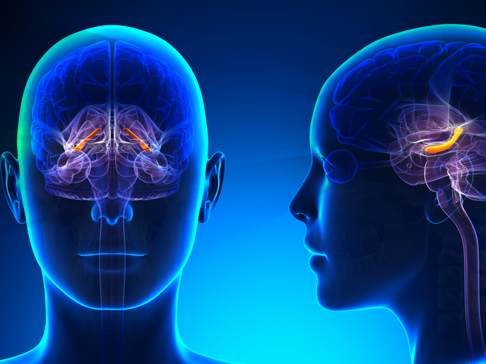
Researchers supported by the EU-funded HBP SGA3 and ICEI projects have created a new high-resolution model of the Cornus Ammonis-1 (CA1) region of a right-side human hippocampus. According to the study published in the journal ‘Nature Computational Science’, this computational method could also be used to generate full-scale scaffold models of other human brain regions starting from microscopy images. The model replicates the structure and architecture of the CA1 region, as well as the position and relative connectivity of the neurons. It was developed from a full-scale data set of high-resolution images that is available in the BigBrain Atlas and will soon be available on EBRAINS, a digital research infrastructure created by the Human Brain Project. “The amount of data on individual neurons of the human brain is very limited, both in terms of relative 3D coordinates and in terms of connectivity among neurons,” comments study senior author Prof. Michele Migliore of HBP SGA3 and ICEI project partner National Research Council, Italy, in a news item posted on the Human Brain Project website. “We have performed a data mining operation on high resolution images of the human hippocampus, obtained from the BigBrain database. The position of individual neurons has been derived from a detailed analysis of these images.”
Finding the neurons
The customised image processing algorithm that has been developed enables a realistic distribution of neuronal positioning. As explained in the news item, an algorithm was also created to generate neuronal connectivity by approximating dendritic and axonal shapes. “Dendrites and axons can be classified into categories depending on the general shape of their extensions: for example, some fit into narrow cones, others have a broad complex extension that can be approximated by dedicated geometrical volumes, and the connectivity to nearby neurons changes accordingly,” states co-first author Dr Daniela Gandolfi from HBP SGA3 and ICEI project partner University of Pavia, Italy. “Our algorithm analyzes high resolution images and, after the creation of specific geometrical shapes to be associated with morphological properties, allows us to calculate the probability that two neurons are connected. The method provides not only the 3D positioning of neurons, but also their connectivity.” To validate their 3D model, the researchers compared the density of neurons in the model with current studies on the hippocampus, and found that they matched. Both the data set and the extraction methodology are on the EBRAINS platform. “Our main goal with this study was to make the data readily available with HBP and the broader neuroscience community. We are now using the same approach to model other brain regions.” HBP SGA3 (Human Brain Project Specific Grant Agreement 3) and ICEI (Interactive Computing E-Infrastructure for the Human Brain Project) end in September 2023. For more information, please see: Human Brain Project website EBRAINS website
Keywords
HBP SGA3, ICEI, Human Brain Project, HBP, human, brain, hippocampus, neuron


