Innovative environment for computer-aided medicine
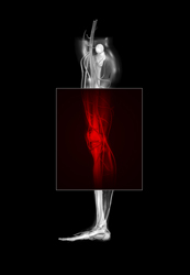
Three-dimensional models of the skeletal muscle structure for the 'typical' patient do not suffice for modern medical practices. Diagnosis and treatment unavoidably rely on the use of non-invasive methods like medical imaging techniques to collect patient-relevant information. Still, surgeons lack tools that would offer a simulated walk through the human body and the option of virtually evaluating new procedures. To address this need, the MULTISENSE project combined haptic devices with visualisation tools in a unique virtual reality environment for orthopaedic surgery. The haptic modality would locate and measure the incision and evaluate the surgical access this incision would provide. In addition, it could help position and orient an implant, as well as, identify any functional impairment produced by soft tissue damage. At the heart of this pre-operative environment for orthopaedic surgery lays a semi-automatic modelling tool that uses data collected from computed tomography (CT) scans to create a patient-specific model. Although CT scans allow for the accurate description of bones under the skin surface, the structure of muscles is difficult to distinguish. Researchers at the Istituti Ortopedici Rizzoli in Italy took up the challenge of designing a dedicated tool that could support the interactive deformation of a generic musculo-skeletal model. This interactive limb anatomy atlas is based on detailed digital representations of the human body that have been developed by the Visible Man project. It employs a simple spring-based model to visualise muscle contraction along with the function of lower limb ligaments. Outlines of skeletal muscles are mapped on this generic musculo-skeletal model and then superimposed on synthetic scans generated along pre-defined axes. These outlines are deformed by simple geometric operations to match the underlying muscle structures to offer an accurate representation of the patient's functional anatomy.


