Scanning innovations target more accurate diagnoses
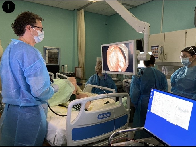
Magnetic resonance imaging (MRI) is a popular technique of capturing images of internal organs and biological systems in the body. Images are generated by using strong magnetic fields and radio waves. A key challenge however is that the human body can be a complex and ‘noisy’ environment to scan. There can be interference in the magnetic field, and patients can have reactions to the contrast agent if one is used. In addition, most MRI machines require the patient to be placed in a narrow tunnel. This can make some people anxious. Speeding up the procedure would therefore improve the patient experience.
New MRI probing and imaging procedures
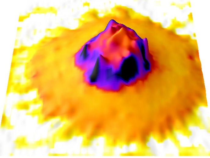
The main goal of the EU-funded PATHOS project was to dramatically improve performance in both MRI and optically detected magnetic resonance (ODMR) sensing. “We began by proposing new theoretical sensing schemes, using both classical and quantum probes for electric and magnetic external fields,” explains PATHOS project coordinator professor Filippo Caruso from the University of Florence in Italy. “Our aim was to drastically reduce the complexity of these probing and imaging procedures. The overarching goal was to substantially enrich the information content extracted from scans and reduce exposure time.” The next step was to develop new scanning techniques capable of achieving the project’s goals, such as through suppressing the thermal ‘noisy’ background in the scan. “We also developed ways of uncovering hitherto invisible biochemical activities,” adds Caruso. The project pioneered new sensing-data processing techniques to exploit all this previously untapped information. “These techniques were then tested using diverse and complementary experimental platforms,” says Caruso. “We also sought to identify future applications that could exploit these new concepts and tools.”
Toolbox to revolutionise biological studies
By going beyond the state of the art in terms of scanning techniques, the PATHOS project was able to create a powerful toolbox. Caruso and his team believe that elements of this toolbox have the potential to revolutionise biological studies, as well as medical diagnostics. In lab-based experiments, many tools were shown to be capable of achieving high levels of scanning accuracy and sensitivity. Some of these experiments involved scanning RNA imino protons, such as those forming the genome of the SARS-CoV-2 virus. This could have implications for the discovery of vaccines and cures. “Some of this fundamental research could open up new opportunities here,” says Caruso. “A good example of this was the application of certain quantum phenomena observed in some experiments. These phenomena were applied to try to better characterise the SARS-CoV-2 virus genome.”
Applying sensing techniques in in vivo environments
Moving forward in the field of diagnostics, Caruso believes that a key next step will be to apply techniques in in vivo environments. “The nuclear magnetic resonance methods developed could be envisaged to work in vivo, as suggested by our results from ex vivo tissue samples,” notes Caruso. “However, the complexity of the chemical environment in live tissue may pose some limits.” Caruso notes that physiological ‘noise’ in a realistic MRI setting could also be challenging to deal with. “These limitations could however be addressed by some of the methods the PATHOS project has proposed,” he adds. “Our hope is that this work will lead to significant time savings, plus enhanced imaging information.”
Keywords
PATHOS, medical, diagnostic, radiation, MRI, biological, magnetic


