

What makes animals move?
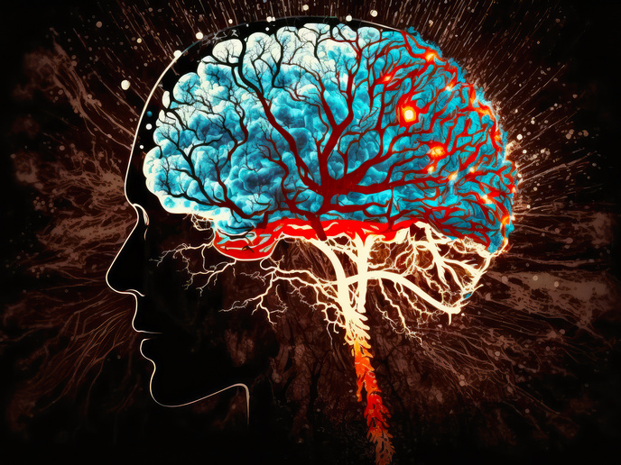
The ability to move is vital for animals to survive in their environment. Researchers supported by the EU-funded EXPLORATOME and ZENITH projects have investigated a particular aspect of movement – forward locomotion – with a focus on the important role played by a brainstem area called the mesencephalic locomotor region (MLR). Published in the journal ‘Nature Neuroscience’, their work has made it possible to map the neuronal circuits involved in initiating forward movement that could help provide valuable insight into movement malfunctions in diseases such as Parkinson’s. Movement can be a conscious process used for navigation. It can also be involuntary, triggered by the senses. “Animals move to explore their environment in search of food, interaction with others, or simply out of curiosity. But the perception of danger or a painful stimulus can also activate an automatic flight reflex,” explains study co-lead author Dr Martin Carbo-Tano of ZENITH project coordinator Paris Brain Institute (ICM), France, in a news item posted on ‘SciTechDaily’. Whether conscious or automatic, movement relies on the activation of command neurons in the brainstem called reticulospinal neurons (RSNs). These neurons relay signals between the brain and spinal cord and play a crucial role in starting, maintaining and stopping movement.
Gaining a global view
The MLR, which triggers forward movement when stimulated, is located upstream of the RSNs. First identified in cats, it is also found in many other vertebrates, such as salamanders, rats, mice, rabbits, guinea pigs, pigs, lampreys and monkeys. “Because the role of the MLR is conserved in many vertebrate species, we assume that it is an ancient region in their evolution—essential for initiating walking, running, flying, or swimming,” adds Dr Carbo-Tano. “But until now, we didn’t know how this region transmits information to the reticulospinal neurons. This prevented us from gaining a global view of the mechanisms that enable the vertebrae to set themselves in motion and, therefore, from pointing out possible anomalies in this fascinating machinery.” To better study the neuronal activity entailed in movement initiation, the research team used zebrafish larvae. The larvae’s transparent brain made it possible for them to identify the MLR and map the downstream command circuits involved in forward locomotion. “We observed that neurons in the mesencephalic locomotor region are stimulated when the animal moves spontaneously, but also in response to a visual stimulus. They project through the pons—the central part of the brain stem—and the medulla to activate a subpopulation of reticulospinal neurons called ‘V2a’. These neurons control the finer details of movement, such as starting, stopping, and changing direction. In a way, they give steering instructions!” remarks study co-senior author Dr Claire Wyart, an ICM researcher and research director at EXPLORATOME project coordinator Institut National de la Santé et de la Recherche Médicale, France. According to the authors, this study supported in part by EXPLORATOME (Circuit mechanisms underlying sensory-evoked navigation) and ZENITH (ZEbrafish Neuroscience Interdisciplinary Training Hub) is an essential step for future research on supraspinal motor control mechanisms. For more information, please see: EXPLORATOME project ZENITH project website
Keywords
EXPLORATOME, ZENITH, animal, neuron, mesencephalic locomotor region, reticulospinal neuron, movement, locomotion, brain