Thirsty rats shed light on our mental autocorrect

Fifteen years ago, neuroscientists discovered a distinctive 6 Hz signal in the brains of people who were on the cusp of making a mistake. Although they could trace it to the mid-frontal cortex, an area of the brain just behind the forehead, it wasn’t clear which neural pathways were responsible for the signal. The EU-funded MidFrontalTheta2.0 project sought to shed light on this poorly understood pattern of brain activity. “When you’re about to make a mistake, for example if you’re typing and you press the wrong key, there is a very specific pattern of electrical activity we can measure,” explains project coordinator Mike X Cohen. “It increasingly bothered me – it was a cool finding and we didn’t know what it meant.”
Brain scans
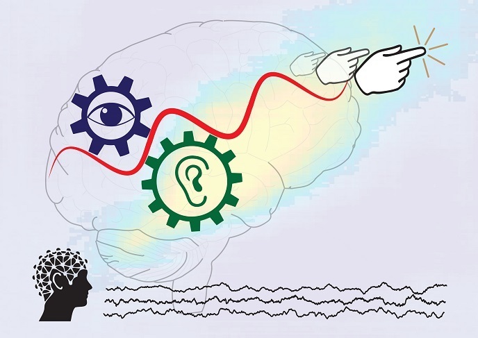
The signal had been measured using an electroencephalogram (EEG) – an array of electrodes attached to the skull which measure electrical activity in the brain. The tool is used extensively in cognitive neuroscience, but can only provide a general idea of brain activity. “We can estimate its location, but that’s not very satisfying – it’s like asking how a car works, and pointing to the front of the car saying ‘the car happens there’,” says Cohen. To gain a deeper understanding of the signal, Cohen and his team decided to work on animal models, which offer ways of recording and manipulating brain activity that is not possible in humans. At Radboud University in Nijmegen, the Netherlands, rats were trained to carry out a task to get water, moving to the left when hearing a high-pitched tone, and to the right when hearing a low-pitched tone. Implanted electrodes simultaneously recorded the front medial cortex and other regions of the rats’ brains.
Crowd noise
With this equipment, the team was able to see fine-grained details in cell populations and circuitry that are not accessible in humans. “When we see the aggregate signal in EEG, it doesn’t tell us how many things are taking place in the brain at the same time,” adds Cohen. “It’s like following a sports game by listening to the noise of the crowd. It’s somewhat meaningful, but there are individuals in that crowd who are cheering for different reasons.” Using the implanted probes and improved data analysis techniques, Cohen and his team were able to see that the signal was generated by multiple brain circuits, all with the same spectral signature. What looked like one signal in EEG was actually multiple synchronous circuits.
High risk
The project was supported by the European Research Council. “This work wouldn’t have been possible without the ERC,” he notes. “This is probably the single biggest risk I ever took in my career.” Cohen credits the success of the project to his team of postdoctorate and PhD researchers. Next, the team plans to work through the data gathered during the last 5 years. “There was no way we were going to be able to understand all this in the timeframe of the funding,” says Cohen. “We’ll spend the next 2 years going through all the data and getting everything published.” He adds that the data generated by the animal experiments will also be made public.
Keywords
MidFrontalTheta2.0, brain, EEG, mistake, front, medial, cortex, electrodes, rats