‘Total heart function’ models offer more effective treatment
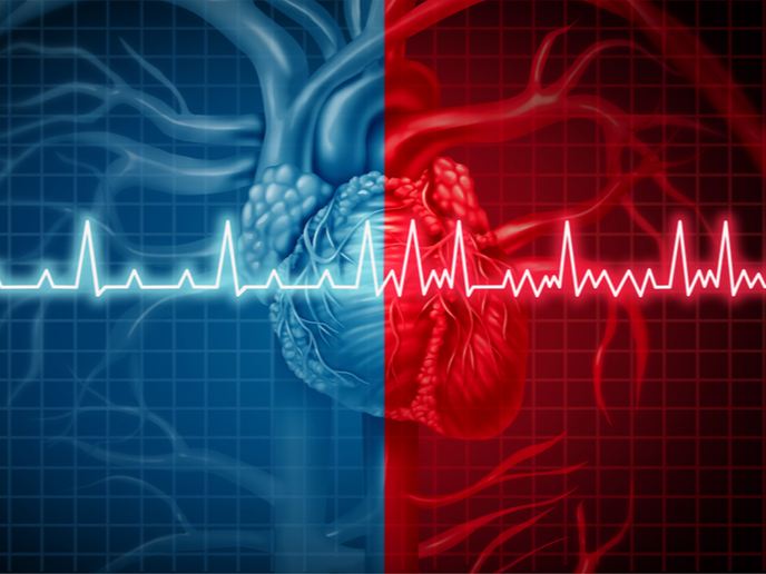
Current tomographic imaging technologies provide a wealth of information about cardiac anatomy, structure and function at a very high, sometimes paracellular, resolution. Yet, personalisation or digital techniques that can fully exploit these rich datasets, and accurately represent patient anatomy and physiology, are still in their infancy. Support from the Marie Skłodowska-Curie programme enabled the InsiliCardio project research fellow to complete a feasibility study into the simulation of personalised total heart function. This was represented in anatomically detailed, high-resolution models, including all three major physics (fluid, structure and electrics). The modelling workflow demonstrated that electro-mechano-fluidic models of the heart are becoming more feasible. While still costly to construct, these models show promise as a tool for predicting response to medical interventions.
Simulating total electro-mechano-fluidic heart function
InsiliCardio combined inputs from wide-ranging disciplines. These included cardiology (cardiac arrhythmias, heart failure and therapies), biomedical engineering (model building, medical image analysis and mapping techniques) and mathematics (numerical methods and scientific computing). This multidisciplinary approach enabled several studies to be conducted that showed the applicability of the computational models for clinically relevant cases. For example, left ventricular wall stresses and biomechanical power are potential biomarkers for diagnosis and prediction of post-treatment outcome. However, these are not accessible within routine clinical procedures or through medical imaging. InsiliCardio modelling allowed these clinically promising biomarkers to be better assessed. Another workstream created atrial (top chambers of the heart) mechanics models, finding that personalised measurements of wall thickness are necessary to accurately calculate local wall stress. Peaks in atrial wall stress are associated with tissue remodelling causing fibrosis – considered a major risk factor for the development of atrial fibrillation. The team developed a simulation framework for investigating this link between local anatomy, mechanics and electrophysiology. “In the short term, our models could improve patient selection, therapeutic planning and risk management, leading to clinical benefits,” says researcher Christoph Augustin. “Specifically, models which can simulate biomarkers that are not available from imaging or clinical measurements, can support decision making in borderline and complex cases.”
Towards better therapeutic planning
Computational models of the heart will impact the lives of European citizens, both as industrial medical device development tools (MDDT), for the design and optimisation of cardiac devices, as well as by offering software as a medical device (SaMD), for diagnostic and therapeutic clinical applications. The design and optimisation of cardiac devices, mechanical heart valves or stents could benefit from the work done by the InsiliCardio project. An open source version of the software called openCARP will be freely available for academic purposes. openCARP currently includes the electrophysiology simulator, with modules for mechanics and fluid dynamics forthcoming. Taking the work forward, Augustin will use InsiliCardio’s data and models within the SICVALVES project, extending them to develop Growth & Remodelling models to further investigate maladaptive hypertrophy (the pathological thickening of a ventricular wall). This is a major risk factor for heart failure. The team’s goal is to help clinicians to plan therapies more effectively. “In the future, we expect that computational models will be employed to assess the state of disease progression and to provide longer-term predictions of therapeutic outcomes,” says Augustin.
Keywords
InsiliCardio, heart, cardiology, cardiac, arrhythmia, ventricular wall stresses, biomechanical, atrial, fibrillation, biomarkers, fibrosis, hypertrophy


