A step towards augmented reality for the treatment of cancer
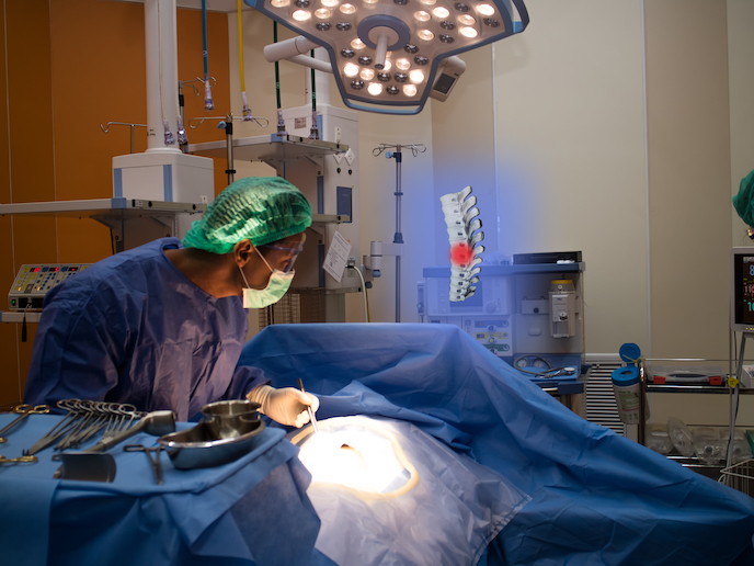
When performing resections, oncology surgeons need tumours to be as clearly defined as possible. Image-guided surgery helps reduce the risk of residual tumour tissue being left behind (resulting in cancer recurrence), while also minimising healthy tissue removal. This approach ultimately reduces patient morbidity and hospital stays, and prevents healthcare costs from spiralling out of control. With the support of the EU’s Marie Skłodowska-Curie programme, the PRISAR project has vastly improved image-guided surgery’s capability. The project developed fluorescence/radionuclide cancer-targeting probes attached to an optical molecule which, when working with a sensitive camera system, can detect vital signals to increase surgical precision and prognosis accuracy. Optical and radioactive detection The technology was developed by first designing a small molecule to target specific cancers. This molecule was then attached to a light-emitting chemical so that when the molecule binds with the cancer, light is emitted which is detectable by a specially adapted camera unit, increasing visibility for the surgeon. A secondary radioactive compound a radioisotope was then constructed to bind to the molecule, once added to the wound bed where the tumour had been excised. The radioisotope is contained locally to the tumour area to minimise radioactive toxicity in other areas of the body. The radioisotope seeks out and destroys any residual cancer cells after surgery, reducing the chances of any remaining cancer cells entering the blood circulation. The project also provided focus for development of a small handheld hybrid detection unit which simultaneously measures both the optical and radioactive signals, avoiding the inconvenience of changing between two specialised cameras. “This is the first camera system in the world that allows multiplexing of fluorescent and gamma probes during the same procedure, demonstrating the potential to take image-guided surgery to the next level, towards augmented reality for medicine,” says molecular imaging specialist Dr Alan Chan. The camera’s modified fluorescent imaging component was originally tested on an immunocompromised mouse, with imaging carried out after the injection of a tracer chemical (111In-DTPA-Trastuzumab-CW800). Clinical imaging was also conducted on a human thyroid gland during a thyroid scintigraphy procedure (gamma scan). Additionally, volunteers undergoing nuclear medicine examinations at Queen’s Medical Centre, Nottingham were imaged to develop the hybrid camera. Towards preventative medicine By increasing the sensitivity of preoperative imaging, when compared to current standards of visual inspection and palpation (physical examination) during surgery, PRISAR’s innovations will benefit patients, oncologists and surgeons. Furthermore, surgeons will be able to use a combination of probes to visualise different physiological and cellular markers, improving the overall sensitivity and specificity of detection. The hybrid gamma-optical imaging device also offers a new tool to tackle existing and emerging medical challenges at the point of care and during intra-operative image guided surgery. “The tangle of anatomical structures, obscured by blood, connective tissue and perhaps even scar tissue, can be challenging to interpret and so any assistance helps reduce errors,” says Dr Chan. “PRISAR’s augmented reality approach can not only identify tumours, but also display their relationship with the surrounding anatomy, including non-diseased tissues.” The team anticipate that this diagnostic toolbox will help pave the way towards more preventative, as opposed to curative, medical approaches. For example, surgery could even be avoided if the camera systems used for image-guided surgery were made sensitive and specific enough to detect early signs of metastases. Currently, it is anticipated that the fluorescent probe will be available in 2022, with the hybrid gamma-optical camera still undergoing clinical evaluation and expected to be market-ready around the same time.
Keywords
PRISAR, fluorescent probe, optical camera, cancer, surgery, metastases, tumour, image-guided, radioactive, tissue, gamma, cell


