Researchers find new ways to interpret modelling systems for diabetes
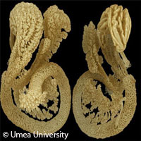
An international team of scientists led by Umeå University in Sweden provides new insight into the aspects of how the pancreas develops during embryonic development and how the so-called islets of Langerhans are distributed in the adult organ. The results of three studies are presented in the journals IEEE Transactions on Medical Imaging, PLoS ONE and Islets; they offer researchers a fresh way to interpret modelling systems for diabetes. The information can be used to fuel our understanding of developmental biology and the adult structure of the pancreas. Researchers use optical projection tomography (OPT) imaging technology to perform three-dimensional (3D) visualizations of gene and protein expression in tissue samples. The drawback of this technology, which can be compared with medical computer tomography, is that it uses ordinary light instead of X-rays; it is commonly used in basic research fields like pathology, plant biology and developmental biology. Scientists at Umeå had already refined OPT in order to perform analyses of entire organs like the pancreas. The information gleaned would help elucidate what goes on with diabetes. In these studies, the team used a novel computer-based technique able to correct distortions in new OPT images. These latest methods give a huge boost to enhanced quantitative and visual OPT analyses in various types of tissues, since sensitivity to small and faintly illuminated objects is heightened. More precise reproductions of the cells depicted in the tissue are also created. Thanks to these novel methods, the researchers shed light on the structure of the pancreas. The islets of Langerhans, found inside the pancreas, produce insulin, the hormone that plays a central role in the regulation of carbohydrates and fat metabolism in the body. When the production of insulin is disrupted and/or when the body's cells fail to respond to insulin signals, diabetes could result. In the study presented in PLoS ONE, the scientists spotlight the developmental biological conditions that lead to the creation of the gastric lobe of the pancreas in the embryo. While the development of this part of the pancreas has not been described thoroughly, the Umeå group identified how this part of the pancreas can only form when the nearby organ, the spleen, develops normally as well. In the Islets-presented study, the scientists identify how the insulin-producing islets are greater in number and are significantly more unevenly distributed in the pancreas than researchers initially thought. They reveal that compared with the rest of the organ, the gastric lobe of the pancreas has a greater number of islets of Langerhans. The findings of these studies provide key information that researchers could use to assess the impact hereditary and environmental factors have on the number of insulin-producing cells in model systems for diabetes.For more information, please visit: Umeå University: http://www.umu.se/english IEEE Transactions on Medical Imaging: http://www.ieee-tmi.org/ PLoS ONE: http://www.plosone.org/home.action Islets: http://www.landesbioscience.com/journals/islets/
Countries
Sweden


