Enhanced imaging of the heart
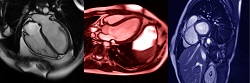
Cardiovascular magnetic resonance (CMR) is a non-invasive imaging procedure aimed at the assessment of the structure and function of the cardiovascular system. The effectiveness of a CMR scan depends on the ability of the operator to correctly tune the acquisition parameters to the subject being scanned and on the potential occurrence of imaging artefacts such as cardiac and respiratory motion. Quality control on the acquired images is usually performed visually by the same operator. To overcome this time-consuming and operator-dependent process, scientists of the EU-funded JUNO project developed an automated image processing approach to improve the quality control of CMR images. “Our ultimate goal was to apply our technique to automatically detect almost in real time incomplete or corrupted scans, allowing the triggering of a new acquisition,″ explains project coordinator Dr Daniel Rueckert. Advancing CMR image processing The JUNO automated technique employs machine learning to extract information from the acquired CMR images. The approach facilitates detection and correction of motion-corrupted images in CMR images by identifying whether the patient could not hold his/her breath as required for proper image acquisition. In addition, it helped validate that the obtained image included the whole heart and also that the cardiac structures were visible and distinguishable. Implementation of the technique on thousands of scans obtained from the UK Biobank demonstrated high accuracy in the detection of problematic scans. With respect to sub-optimal heart coverage detection, the JUNO approach showed 88 % sensitivity and 99 % specificity while slightly lower percentages were obtained for motion-corrupted scan detection. Importantly, the motion correction technique outperformed other existing methods. Clinical implementation of the JUNO technique The JUNO approach is potentially directly applicable in the clinic as a tool for automated quality control of CMR images in the adult. In fact, the relatively low computational time (around 15 seconds are needed to evaluate a typical scan) makes it deployable in the busy clinical routine to inform the operators of potentially corrupted scans and to allow the re-acquisition of the images while the patient is still in the scanner. JUNO researchers believe the technique will also be able to facilitate future clinical studies. With the launch of several initiatives for the acquisition of large-scale open-access databases consisting of thousands of CMR images (such as the UK Biobank), a fully automated quality control pipeline like JUNO is needed to detect corrupted scans and exclude them from the following analyses. Furthermore, the JUNO technique has the potential to be also applied for the motion correction of foetal CMR scans, which are gaining more and more diffusion thanks to recent technological developments. This means that the JUNO approach could provide a more reliable reconstruction of the foetal heart. Although the JUNO approach was not directly tested in the clinic, Dr Rueckert believes “the technique should eventually be released online, freely accessible by research groups and clinics all over the world, addressing the need for reliable fully-automated quality assessment of CMR scans.″
Keywords
JUNO, cardiovascular magnetic resonance (CMR), foetal heart, automated quality control