Super resolution for imaging proteins
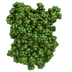
To see when proteins are made and where they go within a cell, scientists use fluorescent molecules that glow when exposed to ultraviolet light. Green fluorescent protein (GFP), originally isolated from a jellyfish that has glowed green for the past 160 million years, is commonly used for this purpose. To make a protein glow green, researchers add GFP to the end of the protein molecule in a process known as tagging. Since the tag can interfere with the protein’s function, the EU-funded GECCCCA (Genetic encoding and click chemistry with copper-chelating azides for super-resolution imaging of proteins) project aimed to develop a new, alternative method to visualise proteins at high resolution under a light microscope. Organic fluorophores are tiny (up to 100 times smaller than GFP) dye-like molecules that can 'stain' proteins by emitting coloured light. Since proteins are made from repeating amino acid units, GECCCCA developed a way to replace a single amino acid within a protein with the dye. They did this by changing the protein’s genetic sequence to include an artificial amino acid that does not exist in nature. They then added the dye to the artificial amino acid using a nature-inspired chemical reaction that joins small molecules together. To test their novel method, GECCCCA stained two highly abundant filamentous proteins that help maintain a cell’s structure (actin and vimentin), with a fluorophore dye. To do this they identified amino acid positions within each protein that could be replaced with an artificial amino acid without affecting the protein’s function. They then tweaked the cell’s protein production machinery so that it could recognise the artificial amino acid, and once produced, linked it to the dye. Lastly, they combined improvements to an imaging system to reveal ultrafine structural detail of individual actin and vimentin filaments in cell compartments. GECCCCA's work will make a significant contribution to the visualisation of proteins in living cells.
Keywords
Proteins, molecular dyes, light microscope, organic fluorophores, artificial amino acid


