Slides that improve optical properties
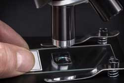
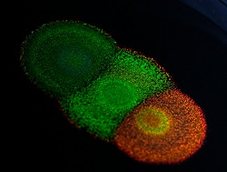
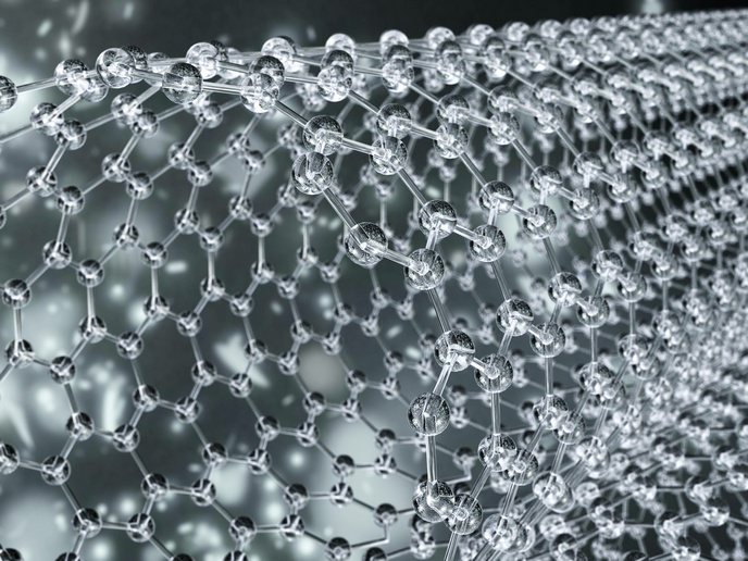
Techniques that support wide-field high resolution, good contrast, and high-data–acquisition rates and signal processing are required for real-time cellular imaging. Important advances in fluorescence-based microscopy have not been able to achieve both high-speed capture of images and high spatial resolution simultaneously for a truly real-time molecular dynamics imaging platform. Fluorescence techniques suffer resolution issues due to the wave nature of light. EU-funded scientists developed biocompatible artificial mesoscopic structures and nanostructures with fundamentally new optical properties. Within the project 'Super-resolution fluorescence microscopy based on artificial mesoscopic structures' (SMARTS), scientists developed materials that can be used as microscopic slides to study live cellular processes. The novel artificial material has been optimised and characterised, including testing for biocompatibility. Nanofabrication methods were demonstrated to be simple and cost effective. Culturing of live cells was straightforward following common protocols and enabled an amazing axial resolution of approximately 10 nm. Further, the advanced technique eliminates the need for scanning, greatly minimising image acquisition and processing time. Results have been published in the prestigious peer-reviewed journal Proceedings of the National Academy of Sciences (PNAS). Researchers exploited their nano-structured material in a study of Förster resonance energy transfer (FRET). This is a fluorescence imaging technique exploiting a donor fluorophore and a receptor fluorophore. It is an excellent sensor at very small distances, but not useful at all for larger distances. The team showed that, through a quantum mechanical effect (the ability of the nanostructure to support surface plasmon modes), their nanostructures can amplify a very low FRET signal. This is a very important finding because most other techniques shown to 'boost' FRET have not been biocompatible. The end of the SMARTS project does not signal the end of the technology development. The team fully plans to incorporate the materials and techniques into a functional imaging platform within the next two years. Collaboration with a prominent lab studying cellular signalling via FRET promises to speed optimisation and commercialisation.
Keywords
Real-time imaging, microscopic slide, mesoscopic structures, nanostructures, fluorescence microscopy


