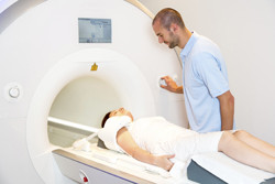
MRI and patient simulator
Since it does not use ionising radiation, MRI is a very safe, non-invasive method to determine both static and dynamic properties of biological tissues. With EU support of the project 'Enhanced MRI physics simulator' (MRISIMUL), scientists have developed a realistic simulation tool that runs on a single computer. The impetus was to integrate realistic aspects of an MRI experiment to understand artefact generation mechanisms during cardiovascular MRI. This will facilitate better exam protocols without the need for more complicated and expensive human or animal experiments. Scientists developed a MATLAB platform that supports development of custom MRI pulse sequences and their application to model objects. MRISIMUL exploits Bloch simulation, the most accurate way to study the effect of pulse sequence on magnetisation. Following analysis of computational power, the team determined that prohibitively long execution times of several days were required even with a high-end personal computer. Researchers replaced the central processor unit (CPU)-based approach with a large number of processing cores integrated with a graphic processing unit (GPU), again on a single computer. Now the computationally demanding core services (kernel) are executed in parallel within the GPU environment for a speed-up of about 228 times compared to serial processing with a CPU. This means, for example, that instead of 5 full days (120 hours), a simulation now requires approximately half an hour. In multi-node, multi-GPU systems, MRISIMUL demonstrated an almost linearly scalable performance with increasing number of available GPU cards. The team has now developed a detailed 3D model of the human heart and torso that simulates relevant motions, including respiration, heart and simple flow. The model can be installed from the project website. There, users will also find a 'fuzzy' spatial distribution of the brain, including 11 tissue types. MRISIMUL has been warmly welcomed by the scientific community with its first description in the Journal of Cardiovascular Magnetic Resonance, acquiring the designation 'highly accessed' within less than a month of its publication. The project also paves the way for extension to other biomedical fields. EU citizens can soon expect better diagnoses and care for a variety of illnesses.
Keywords
MRI, simulator, magnetic resonance imaging, biological tissues, artefact generation, cardiovascular


