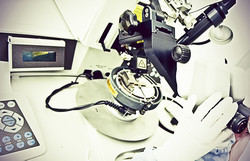
A new multifunctional tool for nanotechnology
The explosion in nanotechnology is having major impact on fields from organic electronics to biomedicine to photovoltaics. The growing volume and diversity of products highlights the pressing need for flexible and multimodal nano-object characterisation technology in a single tool. The EU-funded project UNIVSEM (Universal SEM as a multi-nano-analytical tool) brought together a complementary group of industrial and academic partners whose synergy led to several commercial products. So far, integration of scanning electron microscopy (SEM) and focused ion beam (FIB) with analytical add-ons has been complex. Project partners' activities resulted in several novel developments: integration of a time-of-flight secondary ion mass spectrometer (TOF-SIMS) exploiting the FIB, a high-speed vacuum compatible scanning probe microscope (SPM) with large scan range, and integration of confocal Raman microscope. In addition, new electron detectors, a colour cathodoluminescence detector and a xenon-plasma FIB were developed and integrated, and the overall SEM performance was improved. The first project outcome was a correlative microscopy technique combining SEM and confocal Raman imaging within one integrated microscope system. This combination provides clear advantages for users with regard to comprehensive sample characterisation. SEM is an excellent technique for visualising sample nano-scale surface structures. Confocal Raman imaging is an established spectroscopic method used for detecting a sample's chemical and molecular components. This newly developed microscope enabled for the first time acquisition of SEM and Raman images from the same sample area and correlation of ultra-structural and chemical information. Another project outcome was the successful integration of SEM and SPM with TOF-SIMS, which provides unprecedented analytical capabilities. TOF-SIMS provides better detection limits, higher spatial resolution, depth profiling and the ability to detect isotopic species. Commercialisation is expected to follow closely with major impact on numerous industries thanks to the growing global distribution network of a partner small to medium-sized enterprise. The multimodal tool is foreseen to spur nanotechnology development and enhanced quality control in a myriad of areas, including forensics, geology, biology and optoelectronics.
Keywords
Multifunctional tool, nano-scale analysis, scanning electron microscopy, confocal Raman imaging


