Ultrasound to Combat Liver Tumours
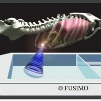
Liver tumours are either benign or malignant; if malignant then they can be primary or secondary. In Europe, a solitary lesion in the liver is more likely to be a metastatic carcinoma than a primary liver tumour. The major risk factors for liver cancer are infection with hepatitis B or C and heavy alcohol consumption, all of which can cause cirrhosis. Smokers and diabetics are also at increased risk, while consumption of foods contaminated with aflatoxin is a cause of liver cancer in many developing countries. Frequently, liver cancer often does not show symptoms until its later stages, so it is seldom diagnosed early. One of the methods for treating a liver tumour is ultrasound. Ultrasound can do much more than record images from the body. Powerful, concentrated ultrasound waves are focused in the patient's body to heat cancer cells to 60 degrees Celsius - destroying them and leaving healthy tissue largely unharmed. Up to now, this 'focused ultrasound therapy' has only been approved for a small number of diseases, such as uterine tumours and prostate cancer. In this context, researchers from the EU-funded FUSIMO project worked on extending the application/method to other organs, such as the liver, which move in the abdomen during breathing. Today, two years after the beginning of the project, many promising intermediate results have been demonstrated. Treating the liver with focused ultrasound presents a major problem: The organ shifts back and forth during breathing. This increases the risk that the ultrasound beam path misses the cancer cells and instead heats the surrounding healthy tissue too strongly. For this reason, researchers have only applied this method to patients under general anaesthesia. To treat a tumour with ultrasound, the medical ventilator is paused for a few seconds so that the patient remains absolutely still. However, general anaesthesia presents its own risks and strains the patient, negating the largest advantage of focused ultrasound therapy - its non-invasive nature. To solve this problem, the FUSIMO project employs a different strategy. If ultrasound therapy applied to a moving liver can be simulated with a computer as closely as possible, the potential for using such treatment on the organ, without general anaesthetic, increases greatly. The ultrasound treatment would either be activated only when the tumour crosses the beam focus, or by tracking the moving abscess so that it remains within the beam path. FUSIMO, coordinated by Fraunhofer MEVIS researchers will develop, implement and validate a multi-level model for moving abdominal organs for use with FUS and magnetic resonance-guided focused ultrasound surgery. Two years on, the project has reached an important milestone: Experts have produced software with which liver operations using ultrasound can be individually simulated for each patient. Magnetic resonance data build the foundation from which 3D images of a patient's abdomen are generated with additional information about the breathing movements over time. Simulations of ultrasound interventions with FUSIMO software are based upon these data sets. To initiate a simulation, researchers enter the time, location, and strength of the desired ultrasound activation. The software created by Fraunhofer MEVIS to efficiently simulate abdominal temperature links two developments: the calculation of ultrasound diffusion provided by the Israeli firm InSightec Ltd. as well as a model of liver movement during breathing from the Computer Vision Lab at ETH Zurich. The software generates an abdominal 'temperature map' that indicates whether a moving tumour has been sufficiently heated and whether the surrounding tissue has been damaged. In the case of suboptimal results, the simulation can be repeated with different parameters. In the long term, the software could help clinicians plan operations and monitor therapy outcomes. At the European Radiologist Congress in Vienna, chief radiologist at La Sapienza University in Rome, Carlo Catalano, stated, "High-intensity focused ultrasound under MRI guidance has become a frequently-applied means of treating non-invasive tumours - for example in the treatment of fibroadenoma of uterus and of bone metastases - but treating tumours in moving organs still represents a major challenge due to several complexities." In this respect, FUSIMO is an exciting project aimed at developing computer simulations for treating the liver with focused ultrasound. In cooperation with both the Institute for Medical Science and Technology (IMSaT) at the University of Dundee and La Sapienza University, MEVIS experts will refine the software during the remaining project year and validate it by comparing experimental data with results from the simulation, which is necessary for determining how realistically the software performs. In principle, this procedure could be applied to other abdominal organs that move with breathing and which are difficult to target with the ultrasound beam path, such as the stomach, kidneys, and duodenum. In addition, specialists are working on a "medicine taxi": cancer medication enclosed in a small fat globule and inserted into the circulatory system. Focused ultrasound beams function as doorkeys to open the globules when inside tumours in organs such as the liver. This process raises the efficacy of the medicine and minimizes harmful side effects. Minimally-invasive procedures help get you out of the hospital and back to daily life sooner than conventional surgery. Because these procedures are less invasive than conventional surgery, there is typically less pain involved and less time spent in the hospital. This novel software will help clinicians plan operations and monitor therapy outcomes, and plan personalised non-invasive highly targeted treatments that will help millions of patients with serious diseases around the globe. FUSIMO stands for 'Patient specific modelling and simulation of focused ultrasound in moving organs.' The project began in 2011 and is funded for three years with 4.7 million euros. Eleven institutions from nine countries are involved. The project is coordinated by the Fraunhofer Institute for Medical Image Computing MEVIS in Bremen, Germany.For more information, please visit: Fraunhofer Institute for Medical Image Computing MEVIShttp://www.mevis.fraunhofer.de/en
Countries
Germany


