Optoacoustic mesoscopy ready to revolutionise medicine
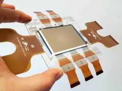
Skin diseases heavily impact society at both socio-economic and healthcare level. Current state-of-the-art optical imaging techniques — such as dermoscopy and confocal microscopy — provide only a partial view of the skin. They are also strongly affected by light scattering, which limits the penetration depth to a superficial few hundred micrometres. These optical techniques cannot visualise the full skin depth, which is around 1.5 mm. Since the skin vascular structure reflects symptoms of a wide variety of diseases from multiple fields of medicine, it would be of great interest and importance to have an imaging method that can visualise the skin vasculature. While ‘Optical coherence tomography’ (OCT) can image slightly deeper than a few hundred micrometres by using coherence gating, the nature of the contrast mechanism does not resolve issues surrounding biologically relevant compounds such as haemoglobin. Similarly, ultrasound can penetrate deep into the tissue, but relies on the administration of external agents to resolve haemoglobin issues at high resolution. With financial support from the EU, the HIFI (Hybrid fluorescence optoacoustic imaging) team assessed the capabilities of a novel optoacoustic mesoscopy system for skin imaging. The outstanding feature of this particular technology is the use of optoacoustic detectors capable of detecting broadband signals. The frequency content of these signals ranges from a few dozen MHz to almost 180 MHz. Such broadband capabilities enable the imaging of objects at different scales, ranging from ~ 5 μm to ~ 100 μm deep in tissue (~4 mm). It is important to note that the system was miniaturised to enable a more user-friendly, handheld operation. Using tissue-like materials, the researchers confirmed the system’s ability to depict small structures identified as the smallest skin capillaries as well as the larger vessels of the deep dermis. In addition, reconstructed images from subsequent in vivo experiments also revealed the whole skin vascular structure plus additional epidermal elements, such as the stratum germinativum and the stratum corneum. The vasculature was derived from the strong optoacoustic signals generated by haemoglobin. This was the first in vivo demonstration of the feasibility of using skin vascular structures for diagnostic purposes. Pilot studies on skin conditions such as psoriasis, eczema, vasculitis and angioma suggested that the system has a tremendous potential to impact the diagnosis of skin diseases and treatment strategies over the long term. Since the skin vascular system indicates symptoms of not only skin-associated diseases but also other malignancies (for example diabetes or hypertension), the impact of optoacoustic mesoscopy is expected to expand well beyond the dermatology field. As a necessary step towards clinical translation of the technology, the HIFI team has already started to move from the proof of concept prototype to building a stable system, exploring the limits of its imaging abilities, identifying specific clinical needs beyond skin-related diseases and quantifying the true impact of the system in the respective clinical setting.
Keywords
Optoacoustic tomography, human tissue, ultrasonic waves, skin diseases, imaging


